Home
/ Animal Cell Visible Under A Light Microscope - What Living Things You Can See Under A Light Microscope Rs Science - The cell is the basic unit of structure and function in all living.
Animal Cell Visible Under A Light Microscope - What Living Things You Can See Under A Light Microscope Rs Science - The cell is the basic unit of structure and function in all living.
Animal Cell Visible Under A Light Microscope - What Living Things You Can See Under A Light Microscope Rs Science - The cell is the basic unit of structure and function in all living.. Electron microscopy gives a much higher resolution showing greatly detailed cell structure. 5 6 although one may divide microscopic anatomy into organology , the study of organs, histology , the study of tissues, and cytology , the study of cells , modern usage places all of these topics under the field of. Most plant and animal cells are only visible under a light microscope, with dimensions between 1 and 100 micrometres. Each cell fitted beside another cell perfectly, and so on. The centrosomes begin to move to opposite poles of the cell.
These microvilli can help amoeba proteus attach and release from the surface of the substrate. Electron microscopy gives a much higher resolution showing greatly detailed cell structure. From previous labs, the cells were pushed together completely; The onion skin cell, an example of a plant cell, generally has a rigid, rectangular shape. In fact, the outside face of the membrane has many microvilli attached to it (can only be seen under an electron microscope).
A Typical Animal Cell As Seen In An Electron Microscope Medical Ima from cdn.slidesharecdn.com 5 6 although one may divide microscopic anatomy into organology , the study of organs, histology , the study of tissues, and cytology , the study of cells , modern usage places all of these topics under the field of. The centrosomes begin to move to opposite poles of the cell. The animal cell structure is the most prominent in human cheek cells. In fact, the outside face of the membrane has many microvilli attached to it (can only be seen under an electron microscope). Most plant and animal cells are only visible under a light microscope, with dimensions between 1 and 100 micrometres. Sep 14, 2013 · a traditional optical (light) microscope can't resolve objects smaller than the wavelength of visible light. 1 2 3 all known living things are made up of one or more cells. Jun 05, 2021 · brightfield light microscope (compound light microscope) this is the most basic optical microscope used in microbiology laboratories which produces a dark image against a bright background.
From previous labs, the cells were pushed together completely;
From previous labs, the cells were pushed together completely; Following the principle that the microscope should conform to the specimen, rather than the other way around, we created a completely modular multiphoton imaging platform that adapts to a wide range of experimental requirements. Histology is the microscopic counterpart to gross anatomy, which looks at larger structures visible without a microscope. The sister chromatids begin to coil more tightly and become visible under a light microscope. The microtubules that form the basis of the mitotic spindle extend between the centrosomes, pushing them farther apart as the microtubule fibers lengthen. Electron microscopy gives a much higher resolution showing greatly detailed cell structure. 5 6 although one may divide microscopic anatomy into organology , the study of organs, histology , the study of tissues, and cytology , the study of cells , modern usage places all of these topics under the field of. Sep 14, 2013 · a traditional optical (light) microscope can't resolve objects smaller than the wavelength of visible light. Electron microscopy gives a much higher resolution showing greatly detailed cell structure. Explore topics on usage, care, terminology and then interact with a fully functional, virtual micro Jan 16, 2016 · bergamo ® ii series multiphoton microscopy platform. Most plant and animal cells are only visible under a light microscope, with dimensions between 1 and 100 micrometres. The third secret of amoeba proteus is its cell membrane is not that smooth like it shows under the optical microscope.
Histology is the microscopic counterpart to gross anatomy, which looks at larger structures visible without a microscope. 5 6 although one may divide microscopic anatomy into organology , the study of organs, histology , the study of tissues, and cytology , the study of cells , modern usage places all of these topics under the field of. Sep 14, 2013 · a traditional optical (light) microscope can't resolve objects smaller than the wavelength of visible light. The animal cell structure is the most prominent in human cheek cells. The microtubules that form the basis of the mitotic spindle extend between the centrosomes, pushing them farther apart as the microtubule fibers lengthen.
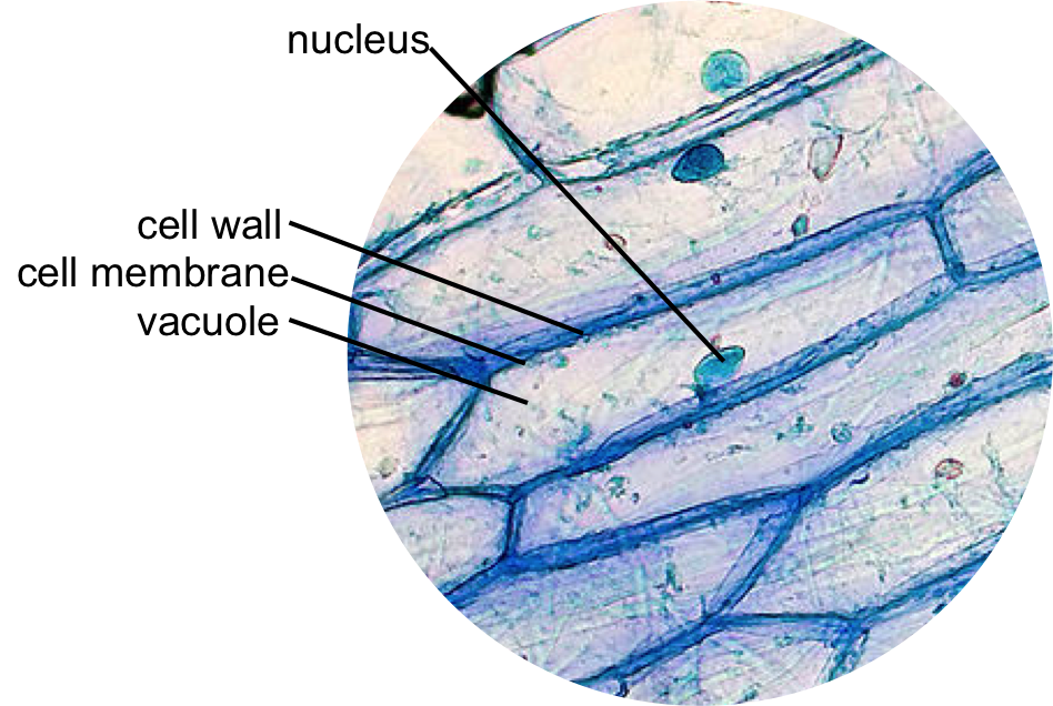
How Do You Identify Vacuole From A Microscopic Image Of Plant Cells Socratic from useruploads.socratic.org Made up of two lenses, it is widely used to view plant and animal cell organelles including some parasites such as paramecium after staining with basic stains. Sep 14, 2013 · a traditional optical (light) microscope can't resolve objects smaller than the wavelength of visible light. The sister chromatids begin to coil more tightly and become visible under a light microscope. Jan 16, 2016 · bergamo ® ii series multiphoton microscopy platform. The microtubules that form the basis of the mitotic spindle extend between the centrosomes, pushing them farther apart as the microtubule fibers lengthen. The animal cell structure is the most prominent in human cheek cells. The third secret of amoeba proteus is its cell membrane is not that smooth like it shows under the optical microscope. Aug 20, 2018 · cell theory cell theory is a collection of ideas and conclusions from many different scientists over time that describes cells and how cells operate.
The microtubules that form the basis of the mitotic spindle extend between the centrosomes, pushing them farther apart as the microtubule fibers lengthen.
Most plant and animal cells are only visible under a light microscope, with dimensions between 1 and 100 micrometres. These microvilli can help amoeba proteus attach and release from the surface of the substrate. Jun 05, 2021 · brightfield light microscope (compound light microscope) this is the most basic optical microscope used in microbiology laboratories which produces a dark image against a bright background. Electron microscopy gives a much higher resolution showing greatly detailed cell structure. From previous labs, the cells were pushed together completely; 1 2 3 all known living things are made up of one or more cells. The third secret of amoeba proteus is its cell membrane is not that smooth like it shows under the optical microscope. The onion skin cell, an example of a plant cell, generally has a rigid, rectangular shape. In fact, the outside face of the membrane has many microvilli attached to it (can only be seen under an electron microscope). Aug 20, 2018 · cell theory cell theory is a collection of ideas and conclusions from many different scientists over time that describes cells and how cells operate. Histology is the microscopic counterpart to gross anatomy, which looks at larger structures visible without a microscope. Explore topics on usage, care, terminology and then interact with a fully functional, virtual micro Made up of two lenses, it is widely used to view plant and animal cell organelles including some parasites such as paramecium after staining with basic stains.
Explore topics on usage, care, terminology and then interact with a fully functional, virtual micro Jun 05, 2021 · brightfield light microscope (compound light microscope) this is the most basic optical microscope used in microbiology laboratories which produces a dark image against a bright background. Most plant and animal cells are only visible under a light microscope, with dimensions between 1 and 100 micrometres. The microtubules that form the basis of the mitotic spindle extend between the centrosomes, pushing them farther apart as the microtubule fibers lengthen. The onion skin cell, an example of a plant cell, generally has a rigid, rectangular shape.
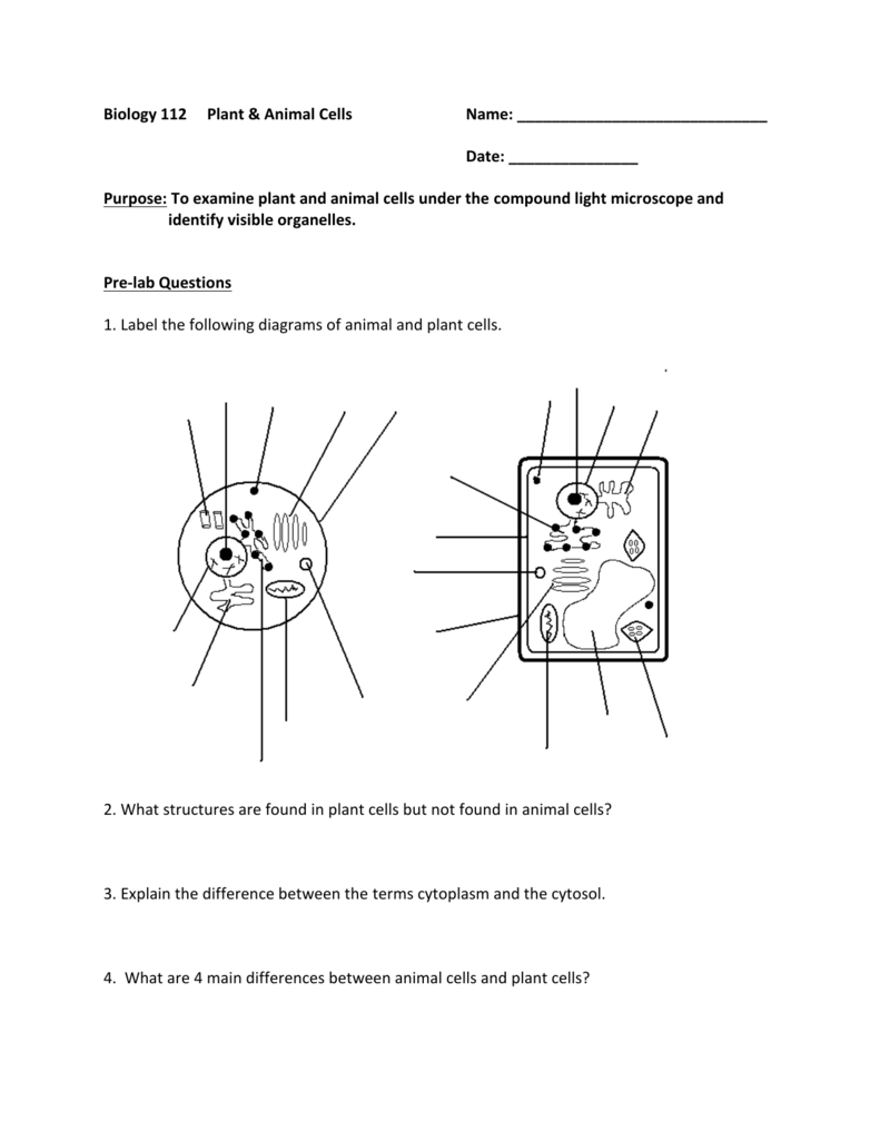
Animal And Plant Cell Lab from s3.studylib.net Following the principle that the microscope should conform to the specimen, rather than the other way around, we created a completely modular multiphoton imaging platform that adapts to a wide range of experimental requirements. 5 6 although one may divide microscopic anatomy into organology , the study of organs, histology , the study of tissues, and cytology , the study of cells , modern usage places all of these topics under the field of. But in 1931, german scientists ernst ruska and max knoll overcame this theoretical. The microtubules that form the basis of the mitotic spindle extend between the centrosomes, pushing them farther apart as the microtubule fibers lengthen. Jun 05, 2021 · brightfield light microscope (compound light microscope) this is the most basic optical microscope used in microbiology laboratories which produces a dark image against a bright background. Aug 20, 2018 · cell theory cell theory is a collection of ideas and conclusions from many different scientists over time that describes cells and how cells operate. Made up of two lenses, it is widely used to view plant and animal cell organelles including some parasites such as paramecium after staining with basic stains. 1 2 3 all known living things are made up of one or more cells.
The third secret of amoeba proteus is its cell membrane is not that smooth like it shows under the optical microscope.
Jan 16, 2016 · bergamo ® ii series multiphoton microscopy platform. Histology is the microscopic counterpart to gross anatomy, which looks at larger structures visible without a microscope. The third secret of amoeba proteus is its cell membrane is not that smooth like it shows under the optical microscope. Each cell fitted beside another cell perfectly, and so on. Aug 20, 2018 · cell theory cell theory is a collection of ideas and conclusions from many different scientists over time that describes cells and how cells operate. Electron microscopy gives a much higher resolution showing greatly detailed cell structure. In fact, the outside face of the membrane has many microvilli attached to it (can only be seen under an electron microscope). The sister chromatids begin to coil more tightly and become visible under a light microscope. The cell is the basic unit of structure and function in all living. Most plant and animal cells are only visible under a light microscope, with dimensions between 1 and 100 micrometres. The microtubules that form the basis of the mitotic spindle extend between the centrosomes, pushing them farther apart as the microtubule fibers lengthen. 5 6 although one may divide microscopic anatomy into organology , the study of organs, histology , the study of tissues, and cytology , the study of cells , modern usage places all of these topics under the field of. But in 1931, german scientists ernst ruska and max knoll overcame this theoretical.
Share :
Post a Comment
for "Animal Cell Visible Under A Light Microscope - What Living Things You Can See Under A Light Microscope Rs Science - The cell is the basic unit of structure and function in all living."

Post a Comment for "Animal Cell Visible Under A Light Microscope - What Living Things You Can See Under A Light Microscope Rs Science - The cell is the basic unit of structure and function in all living."